Korean J Ophthalmol > Volume 23(2); 2009 > Article
Kim, Kim, Kim, and Lew: Serous Retinal Detachment Following Combined Photodynamic Therapy and Intravitreal Bevacizumab Injection

Abstract

We report a case of serous retinal detachment following combined photodynamic therapy (PDT) and intravitreal bevacizumab injection in subfoveal choroidal neovascularization (CNV).
A 53-year-old woman was diagnosed with subfoveal CNV secondary to age-related macular degeneration (AMD) and treated with combined PDT and intravitreal bevacizumab injection. One day after treatment, the patient experienced a sudden decline of vision and optical coherence tomography (OCT) showed serous retinal detachment involving the macula. She was managed conservatively with an oral steroid beginning on the second day of the combined treatment and the subretinal fluid started to decrease one week following the initiation of steroids.
This case suggests that combined PDT and intravitreal injection of bevacizumab can be associated with serous retinal detachment. Additional studies are needed to establish the safety and complications following this treatment regimen.

Intravitreal bevacizumab injection and photodynamic therapy (PDT) with Verteporfin are effective for the treatment of choroidal neovascularization (CNV).1,2 Recently, combined PDT with Verteporfin and intravitreal bevacizumab injection has shown a synergistic effect and might reduce the need for retreatment and cyclic injections of bevacizumab.3
Single treatments of PDT and intravitral bevacizumab injection are considered relatively safe and effective.4,5 However, PDT may cause visual disturbances, subretinal hemorrhage, suprachoroidal hemorrhage and retinal pigment epithelial tear, and there are reported complications following intravitreal bevacizumab injection such as retinal pigment epithelial tear and acute glaucoma.6-11
Chung et al.12 reported a case of retinal detachment with macular hole following combined PDT and intravitreal bevacizumab injection but the safety of combined treatment with these two modalitiesis not well established.
We herein present a case of serous retinal detachment after combined PDT and intravtreal bevacizumab injection in subfoveal CNV.

Case Report

A 53-year-old woman presented with metamorphopsia in her right eye. She was diagnosed with subfoveal CNV secondary to age-related macular degeneration (AMD) and the best-corrected visual acuity of the right eye was 20/50. PDT with Verteporfin was performed according to the standard protocol of the Treatment of Age-Related Macular Degeneration with Photodynamic Therapy Investigation and repeated after 5 months.13 The visual acuity of the involved eye did not change.
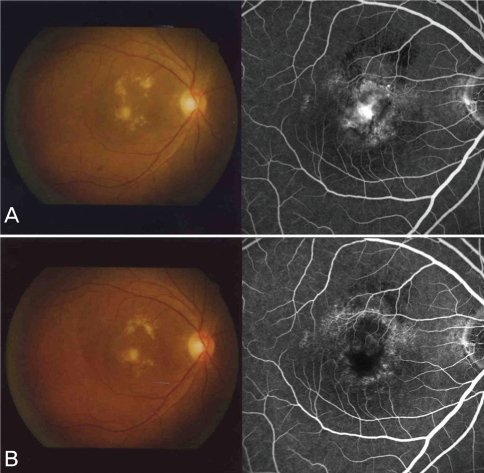
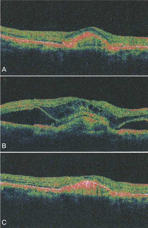
Fifteen months after the second PDT, fluorescein angiography of the affected eye revealed persistent subfoveal CNV and the best corrected visual acuity remained 20/50 (Fig. 1A, 2A). Combined PDT according to the same standard protocol and 1.25 mg intravitreal bevacizumab injection was performed on the same day. One day after the treatment, the patient's vision had decreased to counting fingers. Fundus examination and optical coherence tomography (OCT) showed serous retinal detachment involving the macula (Fig. 2B).
On post-treatmentday two, the patient was initiated on an oral steroid (Triamcinolone® 24 mg #1) to complete a two week course. One week into steroid therapy, the visual acuity remained the same, but OCT findings of the right eye showed decreased serous retinal detachment (Fig. 2C). The vision slightly improved to 20/200 upon completion of the two week steroid course and fluorescein angiography showed decreased leakage of fluorescein dye (Fig. 1B).

Discussion

There have been several reports about serous retinal detachment after PDT treatment.7,9,14 Mennel et al.7 quantified transient serous retinal detachment in CNV after PDT using OCT. Kim et al.9 also suggested that serous retinal detachment might develop following PDT but might also resolve spontaneously several days following treatment. However, there is neither a published report of serous retinal detachment after the combined use of PDT and intravitreal bevacizumab injection nor following intravitreal bevacizumab injection alone.
Several mechanisms may be responsible for subretinal fluid accumulation after combined treatment with PDT and intravitreal bevacizumab injection. The PDT may induce fluid leakage from the CNV itself or damage of retinal pigment epithelium causing an alteration of the blood-retinal barrier.7,9 Bevacizumab may cause instability of the intraretinal and subretinal fluid and subsequent serous retinal detachment by rapidly modulating the permeability and activity of the CNV.10,12
In our patient, there was no evidence of increased subretinal fluid after the first two treatments of PDT alone and the PDT protocol was the same for all three treatments. Occurrence of the serous retinal detachment just after combined PDT and intravitreal bevacizumab injection suggests the interaction of these two modalities as a possible causal role. However, the definitive causation of serous retinal detachment in PDT alone, intravitreal bevacizumab injection alone or combined treatment cannot be determined.
A two week course of oral steroids was used to manage the serous retinal detachment after the combined treatment of PDT and intravitreal bevacizumab injection. The acute inflammatory reaction following either modality may be relieved by systemic steroids. Kim et al.9 injected Triamcinolone® into the vitreous in a patient with serous retinal detachment following PDT, whereas, we elected to use a more conservative management with an oral steroid.
In conclusion, combined PDT and intravitreal bevacizumab injection can be associated with serous retinal detachment which may cause sudden visual loss. Additional studies are needed to establish the standard protocol, safety and complications regarding the combined treatment of PDT and intravitreal bevacizumab injection.

REFERENCES

1. Yoo MH, Boo HD, Kim HK. Result of photodynamic therapy for idiopathic subfoveal choroidal neovascuarization. Korean J Ophthalmol 2005;19:264-268.
crossref pmid
2. Avery RL, Pieramici DJ, Pieramici DJ, et al. Intravitreal bevacizumab (Avastin) for neovascular age-related macular degeneration. Ophthalmology 2006;113:363-372.
crossref pmid
3. Dhalla MS, Shah GK, Blinder KJ, et al. Combined photodynamic therapy with verteporfin and intravitreal bevacizumab for choroidal neovascularization in are-related macular degeneration. Retina 2006;26:988-993.
crossref pmid
4. Fung AE, Rosenfeld PJ, Reichel E. The international intravitreal Bevacizumab safty survey: using the internet to assess drug safety worldwide. Br J Ophthalmol 2006;90:1344-1349.
crossref pmid pmc
5. Rich RM, Rosenfeld PJ, Puliafito CA, et al. Short-term safety and efficacy of intravitreal Bevacizumab (Avastin) for neovascular age-related macular degeneration. Retina 2006;26:495-511.
crossref pmid
6. Schmidt-Erfurth U, Miller JW, Sickenberg M, et al. Photodynamic therapy with verteporfin for choroidal neovascularization caused by age-related macular degeneration: results of retreatments in a phase 1 and 2 study. Arch Ophthalmol 1999;117:1177-1187.
crossref pmid
7. Mennel S, Meyer CH, Eggarter F, Peter S. Transient serous retinal detachment in classic and occult choroidal neovascularization after photodynamic therapy. Am J Ophthalmol 2005;140:758-760.
crossref pmid
8. Gelisken F, Inhoffen W, Partsch M, et al. Retinal pigment epithelial tear after photodynamic therapy for choroidal neovascularization. Am J Ophthalmol 2001;131:518-520.
crossref pmid
9. Kim C, Chung H, Heo JW. Serous retinal detachment in patients with choroidal neovascularization following photodynamic therapy. J Korean Ophthalmol Soc 2007;48:1354-1361.
crossref
10. Shah CP, Hsu J, Garg SJ, et al. Retinal pigment epithelial tear after intravitreal bevacizumab injection. Am J Ophthalmol 2006;142:1070-1072.
crossref pmid
11. Jalil A, Fenerty C, Charles S. Intravitreal bevacizumab (Avastin) causing acute glaucoma: an unreported complication. Eye 2007;21:1541
crossref pmid
12. Chung EJ, Koh HJ. Retinal detachment with macular hole following combined photodynamic therapy and intravitreal bevacizumab injection. Korean J Ophthalmol 2007;21:185-187.
crossref pmid pmc
13. Treatment of age-related macular degeneration with photodynamic therapy (TAP) Study Group. Photodynamic therapy of subfoveal choroidal neovascularization in age-related macular degeneration with verteporfin: One-year results of 2 randomized clinical trials--TAP report. Arch Ophthalmol 1999;117:1329-1345.
crossref pmid
14. Costa RA, Farah ME, Cardillo JA, et al. Immediate indocyanine green angiography and optical coherence tomography evaluation after photodynamic therapy for subfoveal choroidal neovascularization. Retina 2003;23:159-165.
crossref
Fig. 1
(A) Fundoscopic photography and fluorescein angiography showing leakage of fluorescein dye consistent with subfoveal choroidal neovascularization (CNV) before combined photodynamic therapy (PDT) and intravtreal bevacizumab injection. (B) Leakage of fluorescein dye decreased two weeks after the combined PDT and intravtreal bevacizumab injection.
kjo-23-124-g001.jpg
Fig. 2
Optical coherence tomography (OCT) before and after the combined photodynamic therapy (PDT) and intravtreal bevacizumab injection. (A) Before combined PDT and intravtreal bevacizumab injection, OCT image demonstrates a minimally hyporeflective area in the subneuroretinal space. (B) One day after the combined treatment, OCT image reveals significant enlargement of the hyporeflective area, representing the amount of fluid accumulation. (C) One week after taking the oral steroid, the subretinal fluid has absorbed nearly completely, similar to pretreatment OCT findings.
kjo-23-124-g002.jpg


ABOUT
BROWSE ARTICLES
EDITORIAL POLICY
FOR CONTRIBUTORS
Editorial Office
SKY 1004 Building #701
50-1 Jungnim-ro, Jung-gu, Seoul 04508, Korea
Tel: +82-2-583-6520    Fax: +82-2-583-6521    E-mail: kos@ophthalmology.org                

Copyright © 2024 by Korean Ophthalmological Society.

Developed in M2PI

Close layer
prev next