Diabetic Macular Edema Before and After Intravitreal Triamcinolone Injection
Article information
Abstract
Purpose
To compare intravitreal triamcinolone acetonide (IVT) versus natural course in refractory diabetic macular edema.
Methods
In a prospective interventional case series, twenty five eyes with refractory DME which had been allocated to the sham group of a previous clinical trial underwent new examination and optical coherence tomography about 9 months after their first enrollment. Twenty eyes that met the inclusion criteria, visual acuity (VA) < 20/50 and central macular thickness (CMT) > 200 μm, were treated by 4 mg IVT. Evaluations were repeated at 2 and 4 months post-injection to imitate the similar examination intervals after sham injection. Corrected visual acuity and macular thickness changes following IVT were compared to the corresponding changes after sham injection (the natural course).
Results
Visual acuity changes within and between each period were not statistically significant. Visual acuity decreased 0.08 & 0.09 logMAR by 2 months and 0.06 & 0.04 logMAR by 4 months after sham and IVT injections, respectively. The changes of macular thickness after IVT and sham intervention were not meaningful either. However, the difference between thickness changes by 4 months (52±50 μm increase after sham vs. 262±115 μm reduction after IVT) was significant (P=0.014).
Conclusions
Concerning macular thickness, IVT has beneficial effect on refractory diabetic macular edema as opposed to observation. However, considering visual acuity, it does not induce significant difference in comparison to the natural course of the disease.
Macular edema is the most important manifestation of non-proliferative diabetic retinopathy and a predominant cause of legal blindness in diabetic patients. According to the early treatment of diabetic retinopathy study (ETDRS), the treatment of choice for clinically significant macular edema (CSME) is laser therapy. However, some cases of CSME are refractory to laser therapy and do not have a good prognosis with such treatment.1
Recently, some promising results have been shown in different studies for the treatment of refractory diabetic macular edema with intravitreal triamcinolone acetonide (IVT).2-4 Since diabetic macular edema is a chronic disease and the effect of IVT has been shown to be transient,5,6 the comparison of this therapeutic effect with the natural course of this ailment is necessary. In addition, there are a lot of systemic and ocular factors which influence the natural course and the therapeutic response in these patients.6-9 Therefore, controlling of such confounding elements in research programs seems to be essential.
In clinical trials, there is always a possibility of unequal distribution of various characteristics into the allocated groups and adjusting these many factors is rather difficult. Therefore, a study with two types of intervention on one individual group of cases would be able to overcome this problem. We performed a study by IVT injection on patients with refractory diabetic macular edema who had already been observed for a period of time without any treatment. The purpose was to compare the result of IVT treatment with the natural course of diabetic macular edema in one particular group of patients.
Materials and Methods
In this prospective interventional case series study, all of the patients in the control group of a previous clinical trial who had received sham injection (0.1 cc lidocaine 2% subconjunctively) for refractory diabetic macular edema were recalled for examination and reassessment of macular thickness by optical coherence tomography (OCT-2, Zeiss, Dublin, CA) about 9 months after the first presentation. An informed consent was obtained from each patient. Both the primary trial and this part of the study were approved by the Review Board/Ethics Committee of the Ophthalmic Research Centre. The results of the primary trial have not been published yet.
The previous study included eyes meeting criteria for CSME based on the ETDRS definition, with an anticipated unfavorable visual outcome after initial or supplemental macular photocoagulation due to one or more of the following findings: 1- macular ischemia, 2- diffuse macular edema, 3- severe hard exudates (HE) accumulation in the fovea, and 4- lack of response to previous laser photocoagulations with the last one being more than 3 months before. The patients were randomly assigned to the treatment (4 mg IVT) and the placebo groups. This primary trial demonstrated a transient improving effect in terms of visual acuity (VA) and central macular thickness (CMT) in IVT-received eyes versus the placebo-received cases. (Article in press)
In the current study, all of the placebo-received patients in the initial part were recalled for a new clinical and tomographic evaluation. IVT injection was planned for all patients except for those who met the following criteria: (1) intraocular surgery after the first intervention; (2) best corrected VA≥20/50; (3) intraocular pressure (IOP) > 21 mmHg and/or taking anti-glaucoma medication at the time of re-examination; (4) lens opacity severe enough to interfere with VA testing and performing OCT; (5) CMT less than 200 μm measured by OCT; (6) being candidate for intraocular surgery; (7) one-eyed patients; and (8) non-compliance.
Injections were done under sterile conditions in the operating room with topical anesthesia and insertion of a lid speculum. Four milligrams (0.1 cc) triamcinolone acetonide (Kenacort) was injected intravitrealy with a 27-gauge needle through the superotemporal quadrant. IOP was checked about 10 minutes after injection with a Goldman applanation tonometer and anterior chamber paracentesis was performed if IOP exceeded 30 mmHg.
Examinations were performed at 2 and 4 weeks after injection to monitor IOP and the probable complications. In eyes with IOP>21 mmHg, timolol was prescribed twice daily and in refractory cases, one drop of dorzolamide three times a day was added.
Complete ophthalmic examination and OCT were repeated 2 and 4 months after IVT injection in an attempt to replicate the follow-up periods after the primary injections in the first part of the study. The data collected from both the new exams and the evaluations after IVT injection (2nd intervention) were recorded to compare with the results following sham injection (1st intervention).
Ophthalmic examinations, both in the previous trial and in the current study, were carried out by two ophthalmologists who were not masked for the treatment received by the patients. However, best corrected VA measurements and OCT were performed by optometrists who were not aware of the protocols of the two studies. Best corrected VA measurements were based on the Snellen chart and were converted to the logarithm of the minimum angle of resolution (logMAR) scale for statistical evaluations.
Statistical analysis: It was performed using paired t and independent sample t tests for evaluating quantitative variables changes within and between the two interventions respectively. P values<0.05 were considered statistically significant.
Results
The results are presented in two parts: first, the long-term follow up results comparing the findings of the new exams with the previous data in all of the returned patients before the second intervention. Second, the results of IVT injection in which the findings after the two interventions (IVT vs. sham injection) were compared to each other only in cases who met the criteria for IVT administration.
Long-term follow up results: Twenty five cases (16 male, 9 female) of the patients who were in the sham group of the preceding study returned for re-evaluation. The mean time interval between the sham injection and the new examination was 8.9±3.1 months (range: 4 to 15 months).
The comparison of VA changes between the new exam and each previous one (at months 0, 2, and 4 after the sham injection) were not statistically significant which implied on a stable VA in about 9 months of follow up without any treatment (Fig. 1).
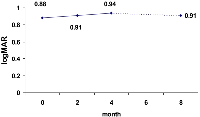
Mean visual acuity before, 2, 4 and about 8 months after the sham injection.
The assessment of CMT between the prior phases and the time of recall are presented in Fig. 2. Although there was a mild increase in mean CMT during the follow up, paired t test did not show statistically significant changes between any of the compared times.

Mean central macular thickness before, 2, 4 and about 8 months after the sham injection.
Results of IVT injection: Five of 25 returning cases were not candidates for IVT injection for the following reasons: VA≥20/50, vitreous hemorrhage, CMT<200 μm (two cases), and non-compliance. Therefore, 20 patients (11 male, 9 female) with the mean age of 62±9 years were treated by IVT. The injections were carried out without any immediate complication except the entrance of drug particles into the anterior chamber in one case due to injection-induced IOP rise.
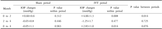
The comparison of VA changes between and within the two interventions (IVT and sham injection) did not show statistically significant difference (Table 1).
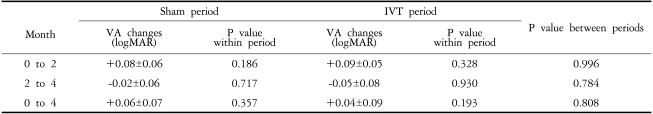
Visual acuity (VA) changes following IVT vs. sham injections in a particular group of cases with diabetic macular edema (paired t and independent samples t tests for changes within and between periods, respectively)
Until 4 months, there was a reduction (-127 μm) in mean CMT subsequent to IVT injection compared to a mild increase (+40 μm) after receiving sham intervention (Fig. 3); however, these differences within each intervention were not statistically significant (paired t test). Comparisons of thickness changes between the two interventions (independent sample t test) did not show significant difference either (P=0.119 for month 0 to 2, and P=0.968 for month 2 to 4), except in one stage. As shown in Table 2, the difference of changes between months 0 and 4 was significant (P=0.014), which implied on CMT reduction after IVT treatment in eyes which had been followed without any treatment for 9 months.
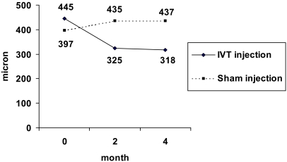
Mean central macular thickness changes after sham and IVT injections.
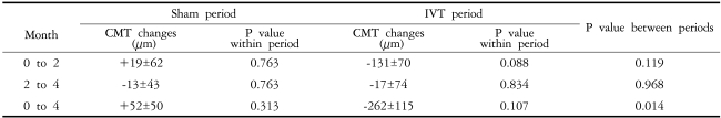
Central macular thickness (CMT) changes following IVT vs. sham injection in a particular group of cases with diabetic macular edema (paired t and independent samples t tests for changes within and between periods, respectively)
Paired t-test showed that IOP rise after receiving IVT was significant at months 2 and 4 (Fig. 4). Although mean IOP before IVT injection was 1.25 mmHg higher than it was before the sham treatment, the comparison of IOP changes following the two interventions demonstrated a significant increase of pressure with triamcinolone especially by 2 months (Table 3).
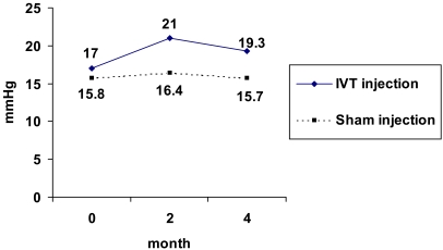
Mean intraocular pressure changes after sham and IVT injections.
Intraocular pressure (IOP) changes following IVT vs. sham injection in a particular group of cases with diabetic macular edema (paired t and independent samples t tests for changes within and between periods, respectively)
Discussion
According to the results, VA and CMT in the patients with refractory diabetic macular edema did not undergo significant changes during 9 months of follow up. IVT treatment in such eyes reduced CMT by 4 months; however, its therapeutic effect on VA was not significant.
We could not detect any statistically significant VA improving effect after IVT injection, neither by before-and-after comparison nor in relation to the changes following sham injection. Jonas et al. showed that in 68% of IVT-received eyes VA improved for 2 Snellen lines; however, in control eyes the VA remained unchanged for 4 months and then it decreased until the end of the follow up. The difference of VA changes between the two groups in their study was significant.10 Note that the dosage of IVT in that study (25 mg) was higher than that of ours, and their control group underwent focal or grid macular laser therapy which indeed could have effects on the result.
Parolini demonstrated a transient VA improvement during 3 months after IVT treatment which was followed by a deterioration of VA until 1 year.9 Considering the effect of IVT on VA in refractory diabetic macular edema, different authors have mentioned different results. For instance, significant increase of VA by 6 months was reported in one study;11 in another, the authors did not find any considerable difference between control and case groups with 24 months of follow up.8
Comparison of CMT changes following IVT injection with its natural course in the present study demonstrated the improving effect of this treatment on macular edema by 4 months. We did not perform OCT after 4 months; however, most of the similar studies with longer follow ups showed that the effect of IVT on macular thickness was transient.6-9 In a study on Chinese patients with diabetic macular edema, 4 mg IVT reduced CMT significantly from 552±179 μm to 326±145 μm and 427±145 μm at months 3 and 6 respectively.6 In Krepler's study with 9 months of follow up, thickness reduction (from 450±190 to 305±153 μm) was significant only at month one.7
It is well known that IVT increases IOP which in most cases can be controlled by anti-glaucoma drugs. In a study by Jonas on 272 patients with different diseases treated by 20 mg IVT, IOP more than 21, 30, 35, and 40 mmHg occurred in 112 (41.2%), 31 (11.4%), 15 (5.5%), and 5 (1.8%) respectively. These IOP rises were treated by medication in all except 3 cases.12 Although this complication is treatable, it may persist beyond the therapeutic effect of the drug. IOP elevation lasted for 8 months in the studies conducted by Jonas12 and Martidis.4
As we found from literature review, no study had been conducted to compare the effect of IVT on refractory diabetic macular edema with its natural course in one particular group of patients. The chronic nature of diabetic macular edema gave the opportunity for planning such a research. Since multiple and different ocular and systemic factors could affect the course of this disease and its response to any treatment modality, the present study with two kinds of managements on one group of cases probably had the power to ameliorate the influences of these confounding elements. However, there could have been some changes in the patient's systemic status between the times of two interventions that we were not aware of them. This fact along with rather small sample size can be regarded as limitations of this study.
In summary, this study like many others implied on the beneficial effect of IVT on diabetic macular edema. However, this effect was evident on macular thickness rather than on VA. Such discrepancy in responses of variables was seen also in Larsson's study.13 Considering the stable nature of VA and macular thickness in patients with refractory diabetic macular edema, and the insignificant effect of IVT on VA, we would not recommend IVT as a routine treatment modality for such cases.
Notes
* Supported by the Ophthalmic Research Center of Shaheed Beheshti University of Medical Sciences