Peripheral Lattice Degeneration Imaging with Ultra-Widefield Swept-Source Optical Coherence Tomography
Article information
Abstract
Purpose
To investigate a series of peripheral lattice degeneration cases using an ultra-widefield (UWF) swept-source optical coherence tomography (SS-OCT) system.
Methods
From August 1, 2022 to July 31, 2023, 19 eyes from 16 patients with peripheral lattice degeneration were included. They all underwent a UWF SS-OCT examination. Anatomy of retina, vitreous, and associated pathologic changes were assessed.
Results
UWF SS-OCT showed various anatomical changes of retina and vitreous in patients with lattice degeneration. Of 15 eyes from 12 patients whose UWF SS-OCT images were clearly obtained, eight eyes showed regional retinal thinning, seven eyes showed vitreous traction, two eyes showed detached vitreous, and three eyes showed retinal break.
Conclusions
UWF SS-OCT can be a useful tool to understand anatomical changes and pathophysiology of peripheral lattice degeneration.
Lattice degeneration is the most common type of retinal degeneration that happens especially in the peripheral retina. Its prevalence is about 6% to 8% of the general population. It more frequently happens in myopic eyes [1–3]. There was no sexual or racial difference in its prevalence, and about 50% of those who have lattice degeneration have it bilateral [4]. In fundus examination, lattice degeneration is characterized by sharply demarcated, circumferentially oriented, oval or round areas of retinal thinning with overlying vitreous liquefaction, and exaggerated vitreoretinal attachments along its edges [5]. Lattice degeneration is clinically important because it can lead to retinal break and even detachment. Due to the risk of retinal detachment, prophylactic treatment is usually performed, including barrier laser photocoagulation and cryopexy for lattice degenerations with retinal breaks or myopic, aphakic, and pseudophakic eye, and past history of retinal detachment on the other eye. However, indication of prophylactic treatment has been ambiguous [6–9]. Some studies have visualized and analyzed anatomic changes of lattice degeneration and adjacent vitreous using spectral domain optical coherence tomography (SD-OCT) and OCT angiography. However, these devices can only capture central retina, peripheral lattice degeneration could not be characterized [3,10–13]. Ultra-widefield (UWF) swept-source OCT (SS-OCT) system is a novel device that combines UWF scanning laser opthalmoscope (SLO) with a navigated SS-OCT (Silverstone, Nikon Healthcare Japan Inc). It can produce an initial high-resolution UWF SLO fundus image of up to 200°. It uses navigated SS-OCT line or volume scans at any location as mapped by UWF SLO fundus images, making it possible to show tomographic images in the peripheral retina [14,15]. In this paper, anatomy of peripheral lattice degeneration and changes of retina and vitreous following lattice degeneration using UWF SS-OCT system were analyzed.
Materials and Methods
Ethics statement
The study was approved by the Institutional Review Board of Soonchunhyang University Seoul Hospital (No. 2023-04-017). The study protocol adhered to the tenets of the Declaration of Helsinki. The requirement for informed consent was waived due to the retrospective nature of the study
Study design and setting
This is a retrospective study that reviewed medical records of patients diagnosed with peripheral lattice degeneration and underwent UWF SS-OCT in Soonchunhyang University Seoul Hospital (Seoul, Korea) during the period of August 1, 2022, to July 31, 2023.
A total of 97 patients who suffered from floater or had a history of retinal detachment were the subject. Of these 97 patients, 19 eyes from 16 patients who had peripheral lattice degeneration were included regardless of the presence of previous laser scar. Lattice degeneration was diagnosed through UWF photos and fundus examination by clinicians under dilation. Of 19 eyes with lattice degeneration, four eyes were excluded because authors were not able to obtain clear UWF SS-OCT images. For two eyes, lattice degenerations were located in far periphery and UWF SS-OCT could not capture them in primary gaze. For the other two eyes, UWF SS-OCT captured lattice degenerations but the obtained image qualities were too poor to analyze. All subjects underwent comprehensive ophthalmic examinations including best-corrected visual acuity, intraocular pressure, slit-lamp examinations, medically induced mydriasis, fundus examinations, UWF SS-OCT for UWF fundus imaging, and OCT imaging. All UWF SS-OCT imaging procedures were conducted by trained clinicians using UWF high definition 6-mm volume cross-sectional scan mode on the UWF fundus photograph obtaining 121 images for each trial. Obtained images were diagnosed and analyzed by two clinicians (KSC and JK). We defined peripheral lattice degeneration as lattice degenerations located beyond the major vascular arcades. SS-OCT images of peripheral lattice degeneration were analyzed, especially focusing on the anatomical changes of retina, relation between vitreous and lattice degeneration including findings like subretinal fluid, retinal break, vitreoretinal traction, and detached vitreous.
Results
Fifteen eyes from 12 patients (five men and seven women) who had peripheral lattice degeneration and underwent UWF SS-OCT imaging were included. Basic demographics of subjects are shown in Table 1. Fifteen OCT images were reviewed thoroughly and classified into five distinct anatomical changes of the lattice degeneration and adjacent vitreous around lattice degeneration. Retinal thinning in eight eyes, retinal break without vitreous traction in one eye, vitreous traction in seven eyes, retinal break with vitreous traction in two eyes, and detached vitreous from underlying lattice degeneration in two eyes were observed. The UWF fundus images of lattice degeneration including guideline for UWF SS-OCT image and cross-sectional SS-OCT images are demonstrated in Fig. 1–5. Various findings are described as follows (Table 2).

Basic demographic characteristics of patients
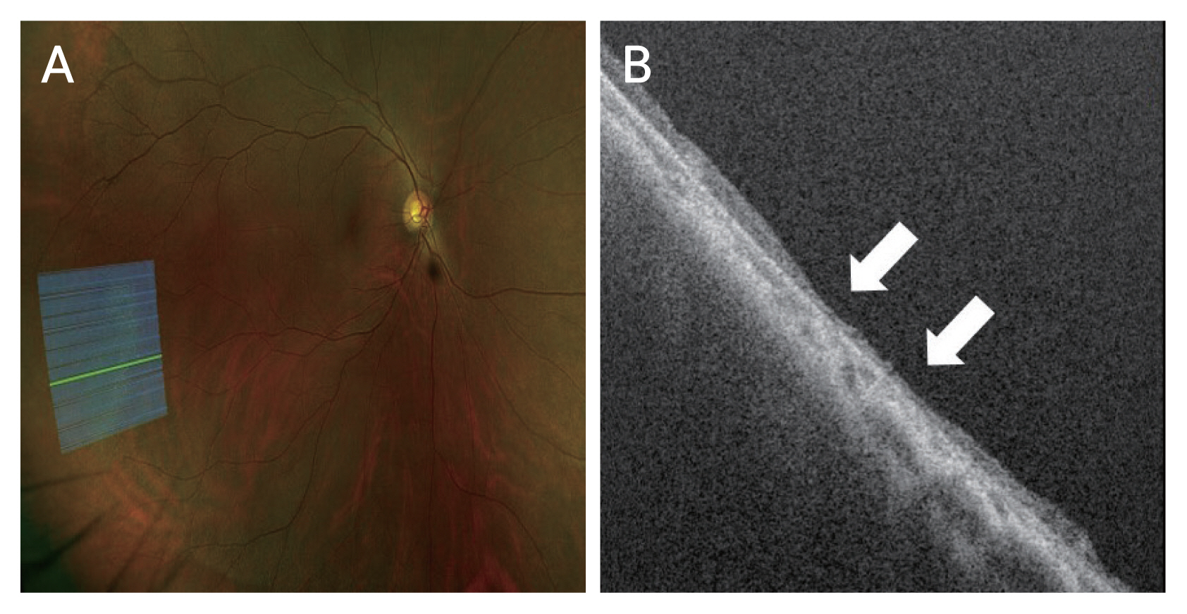
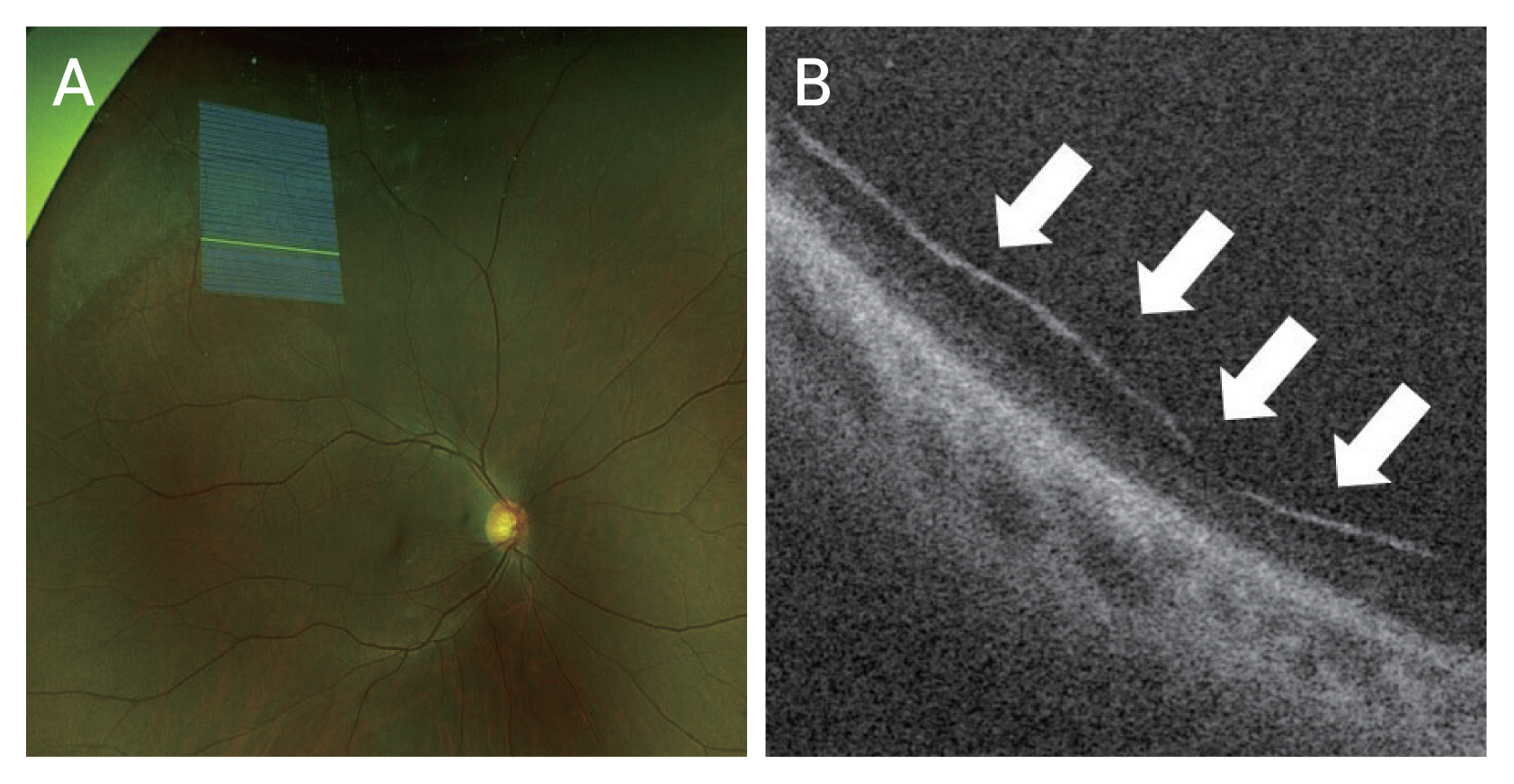
Retinal thinning. (A) Ultra-widefield (UWF) fundus photograph of lattice degeneration and guideline to swept-source optical coherence tomography (SS-OCT). (B) UWF SS-OCT image corresponding to the line of UWF fundus photography. UWF SS-OCT image shows generalized thinning and disruption of division of retinal layers (arrows).
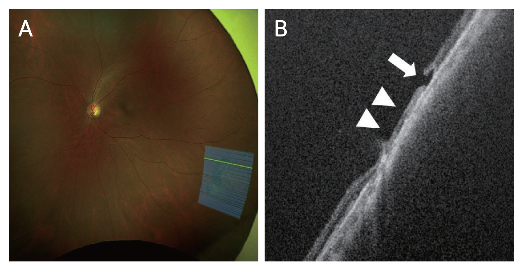
Retinal break. (A) Ultra-widefield (UWF) fundus photograph of lattice degeneration and guideline to swept-source optical coherence tomography (SS-OCT). (B) UWF SS-OCT image corresponding to the line of UWF fundus photography. UWF SS-OCT image shows retinal break that is not obvious in the fundus photography (arrow) along with retinal thinning (arrowheads).
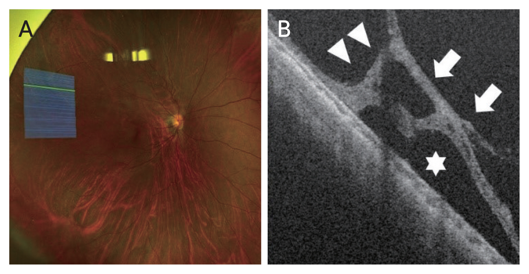
Vitreous traction. (A) Ultra-widefield (UWF) fundus photograph of lattice degeneration and guideline to swept-source optical coherence tomography (SS-OCT). (B) UWF SS-OCT image corresponding to the line of UWF fundus photography. UWF SS-OCT image shows vitreous traction (arrows) with localized retinal detachment (star) and retinoschisis (arrowheads).
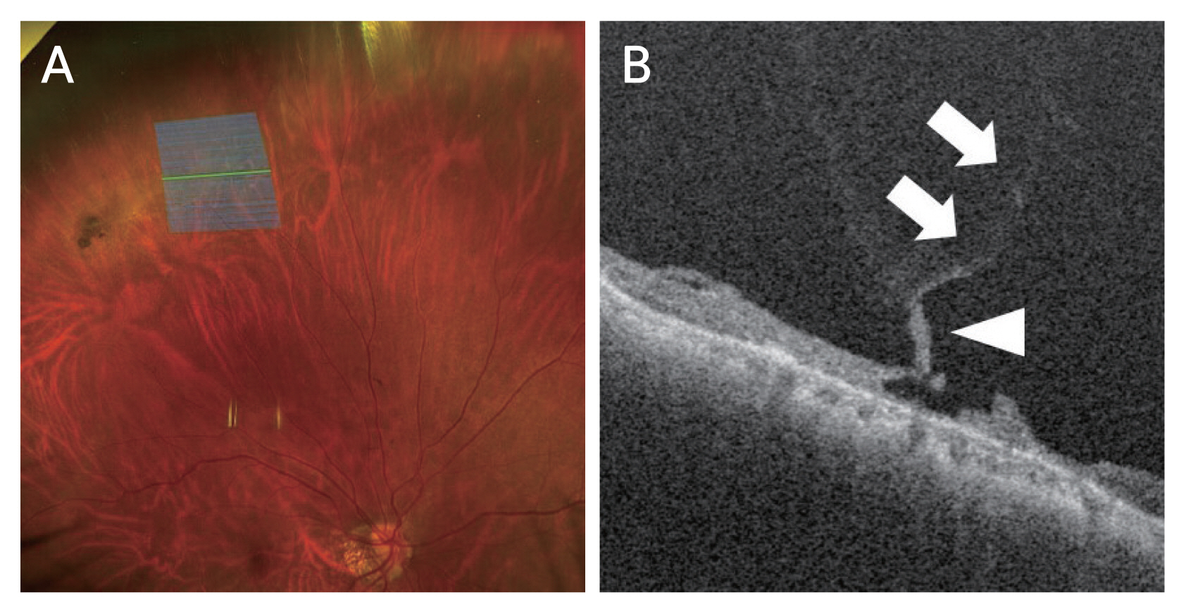
Vitreous traction with retinal break. (A) Ultra-widefield (UWF) fundus photograph of lattice degeneration and guideline to swept-source optical coherence tomography (SS-OCT). (B) UWF SS-OCT image corresponding to the line of UWF fundus photography. In UWF-SSOCT image, a vitreoretinal traction is observed (arrows), retinal flap is seen connected to the vitreous (arrowhead).
Detached vitreous membrane. (A) Ultra-widefield (UWF) fundus photograph of lattice degeneration and guideline to swept-source optical coherence tomography (SS-OCT). (B) UWF SS-OCT image corresponding to the line of UWF fundus photography. UWF SS-OCT image shows a detached vitreous membrane (arrows).

Findings of lattice degeneration (n = 15)
Retinal thinning
The most common finding was retinal thinning. Of eight eyes (53.3%), retinal thinning was found in lattice degeneration (Fig. 1A, 1B). There was overall thinning of retinal layer having ambiguous border of inner and outer retina. Retinal layer thickness was obviously thinner than surrounding normal tissues.
Retinal break
In one eye (6.7%), retinal break was found in lattice degeneration (Fig. 2A, 2B). Retinal break was not clearly seen in fundus photograph, but obvious in OCT scan images. There was retinal thinning around the retinal break.
Vitreous traction
In seven eyes (46.7%), vitreous traction was found in lattice degeneration (Fig. 3A, 3B). In all cases, retinoschisis and focal retinal detachment were found as well. Vitreous strand was obviously attached to the retina and pulled it anteriorly, making different degrees of retinoschisis.
Vitreous traction with retinal break
In two of seven eyes (13.3%) that showed vitreous traction, there were retinal breaks (Fig. 4A, 4B). There was a retinal break with an operculum connected to the vitreous strand above it.
Detached vitreous membrane
In two eyes (13.3%), detached vitreous membrane above the lattice degeneration was observed (Fig. 5A, 5B). A long strand of vitreous membrane was above the retinal layer, showing obvious gap between the vitreous and lattice degeneration.
Discussion
Since the development of OCT, we can understand the anatomy of retina and pathological changes of retinal layers in various diseases much better than before. OCT gives us high-resolution images of retina, choroid, and vitreous with a noninvasive method. In clinics, OCT examination is one of the most important examinations. Many clinicians make decisions depending on results of OCT examination [11,12,16]. However, traditional OCT could not capture pathologic changes outside of major vascular arcades of fundus. Lattice degeneration is a well-known disease usually found in peripheral retina in fundus examination. The reason why it is important is because lattice degeneration is related to retinal breaks and detachment. Histological findings of lattice degeneration including vitreous is firmly attached to the margin of lattice degeneration and that vitreous is liquified over the lattice degeneration are well-known [2]. However, because it is usually located in peripheral retina, it was hard to perform imaging study with OCT. In the current study, we were able to capture OCT images of lattice degeneration located in peripheral retina using UWF SS-OCT. We investigated 15 OCT volume scan images of lattice degeneration. With UWF SS-OCT, we were able to capture peripheral lattice degeneration and the vitreous around it as well as the relation between them with cross-sectional images. The most common change of peripheral lattice degeneration was retinal thinning. Compared to adjacent normal peripheral retina, the border of inner and outer retina was ambiguous, showing homogenous reflectivity through all layers. Retinal breaks with and without vitreous traction were found as well. And there was a retinal break in lattice degeneration that was hardly seen in fundus photograph. In a previous study [13] that analyzed OCT images of posterior lattice degeneration, 46% of the studied eyes showed U-shaped vitreous traction, 23% eyes showed retinal thinning, 15% eyes showed vitreous membrane and retinal break. In this study, vitreous membrane, vitreous traction, and retinal break showed similar incidence. However, retinal thinning was found in 53.3% of studied eyes, which is much higher than the previous study. This difference is maybe due to the small number of patients in both studies. Or the fact that this study focused on peripheral lattice degeneration compared to the previous study that conducted in patients with posterior lattice degeneration. Because of the low incidence of retinal detachments due to lattice degenerations shown in many studies, prophylactic treatment of lattice degeneration was controversial [6,7,17]. For many clinicians, lattice degeneration alone was not an indication of barrier laser photocoagulation if there were no retinal breaks, previous history of retinal detachment, or family history of retinal detachment [4]. However, with UWF SS-OCT, we can now detect retinal breaks in lattice degeneration that can hardly be detected in fundus examination. We can also differentiate lattice degenerations with vitreous traction from lattice degenerations without vitreous traction. Those lattice degenerations alone could be the subject of barrier laser photocoagulation.
Limitations of this study include its retrospective design and relatively small number of patients included. UWF SS-OCT can capture OCT images of lattice degeneration only when patients are looking straight ahead, not tilting their eyes. Thus, peripheral lattice degenerations captured in UWF fundus photos by tilting patient’s eyes could not be included. In addition, there were some UWF SS-OCT images that were too poor to analyze even though they were captured in UWF fundus photos in primary gaze.
In conclusion, we could capture OCT images of peripheral lattice degeneration that were impossible with traditional OCT using a UWF SS-OCT system even though there were some limitations. By analyzing OCT images, we could understand the anatomy of peripheral lattice degeneration and the vitreous around it better. We were able to detect retinal breaks in lattice degeneration that was not obvious in fundus photograph alone. We found that if there was vitreous traction above peripheral lattice degenerations, barrier laser photocoagulation should be considered to prevent retinal break and detachment even though there were no retinal breaks.
Acknowledgements
None.
Notes
Conflicts of Interest
None.
Funding
This study was supported by a Soonchunhyang University Research Fund. The funder had no role in case selection, decision to publish, or preparation of the manuscript.