Bilateral Acute Angle-Closure Glaucoma after Macular Hole Surgery
Article information
Dear Editor,
Face-down positioning after macular hole surgery is routine. We describe a case of bilateral acute angle-closure glaucoma (AACG) as a complication of face-down positioning after macular hole surgery.
A 65-year-old Korean female visited our clinic with a chief complaint of right visual disturbance lasting several months. On initial examination, the best-corrected visual acuity was 20 / 200 (oculus dexter, OD) and 20 / 20 (oculus sinister, OS). The intraocular pressure (IOP) as measured by Goldmann-applanation tonometry was 13 mmHg (OD) and 14 mmHg (OS). A slit-lamp examination revealed a shallow anterior chamber and moderate nuclear sclerotic cataract on both eyes. No glaucomatous optic neuropathy was evident on either eye, and a full-thickness macular hole was identified in the right eye on fundus examination. The patient underwent a pars plana vitrectomy with internal limiting membrane peeling and C3F8 gas tamponade. Postoperatively, the patient was positioned face down. On day 1, the IOP was 13 mmHg in both eyes, and topical treatment with moxifloxacin hydrochloride (Vigamox; Alcon Laboratories, Fort Worth, TX, USA), 1% prednisolone acetate (Pred-forte; Allergan plc., Dublin, Ireland) and 1% atropine sulfate (Isopto Atropine, Alcon Laboratories) was started.
On the 7th postoperative day, the patient complained of visual disturbance and ocular pain in both eyes. Her visual acuity had dropped to hand motion in both eyes. On slit-lamp examination, shallow anterior chambers with occluded angles (Shaffer grade 0) and mid-dilated fixed pupils were observed (Fig. 1A). The IOPs were 41 mmHg (OD) and 40 mmHg (OS) by Goldmann-applanation tonometry. Under the diagnosis of bilateral AACG, the patient received systemic treatment with intravenous 15% mannitol and 500 mg oral acetazolamide hydrochloride as well as topical therapy with timolol-dorzolamide fixed combination (Cosopt; Merck & Co., Kenilworth, NJ, USA), brimonidine (Alphagan P, Allergan plc.), tafluprost (Taflotan; Santen Pharmaceutical, Osaka, Japan) and 2% pilocarpine (Isopto Carpine, Alcon Laboratories), and the IOP decreased to 22 mmHg (OD) and 12 mmHg (OS). After IOP and pain control with medical therapy, laser peripheral iridotomy with concomitant laser peripheral iridoplasty on both eyes was performed. After cessation of all medical treatment, the anterior chamber deepened, and the IOP remained within normal limits. Gonioscopy performed 2 weeks after laser iridotomy predominantly revealed a grade 1 angle width (Schaffer), but partial iridotrabecular contact was observed in both eyes (Fig. 1B). The best-corrected visual acuity was 20 / 60 (OD) and 20 / 25 (OS) with normal IOP ranges. The axial lengths were measured at 21.57 mm (OD) and 21.50 mm (OS). However, the partial iridotrabecular contacts remained and glaucomatous optic neuropathies with visual field defects were evident in both eyes, so cataract surgery was performed. Postoperatively, the depth of the anterior chamber increased and the IOP stabilized (Fig. 1C).
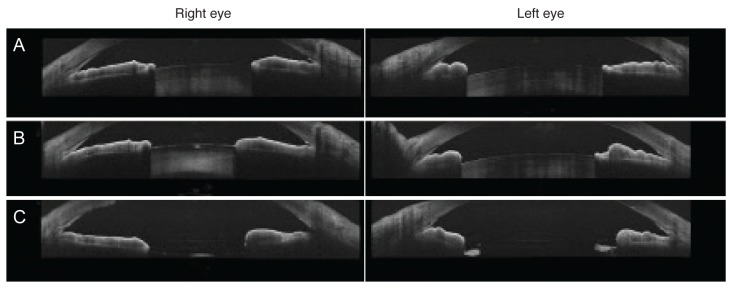
Anterior-segment optical coherence tomography image of the patient. (A) Before laser iridotmy, acute angle-closure glaucoma showed bilateral occluded angle. (B) After laser iridotomy, the angle widened, but partial iridotrabecular contact remained. (C) After lens extraction, the remaining iridotrabecular contact was resolved. The authors received written permission from the patient to report the case.
Face-down positioning (to provoke angle closure and detect dramatic rises in intraocular pressure) has been suggested as a method to identify those at high risk of angle closure. The prone position induces forward movement of the lens and enhances the effect of relative pupillary block [1]. Face-down positioning after macular hole surgery is effectively an extended prone-position provocation test.
Several previous case reports identified prolonged prone or face-down positioning as a cause of AACG. Those cases were bilateral, and they occurred after prone positioning during spinal surgery on patients without any previous ophthalmologic pathology or surgical history [23]. Sutter et al. [4] reported a case of AACG in the fellow eye during the postoperative phase of retinal detachment repair. The AACG occurred only in the fellow eye because the eye that had undergone surgery was pseudophakic, leaving the anterior segment less crowded and filled with lighter-than-water silicone oil, thus relieving any possible pupillary block. Even though the operated eye had been gas-tamponaded in the present case, the AACG was bilateral. In both Sutter et al. [4] (22.79 mm) and the present case, the axial length of the AACG eye was relatively short.
Face-down positioning poses a risk of AACG in predisposed patients. Surgeons should be aware of this complication, as timely diagnosis and treatment are required to prevent visual deterioration. We encourage our colleagues to assess AACG risk in both eyes during the preoperative evaluation (including gonioscopy), especially for at-risk patients. If occludable angles are found, prophylactic treatment with laser peripheral iridotomy appears to be of value in preventing a postoperative IOP spike.
Notes
Conflict of Interest: No potential conflict of interest relevant to this article was reported.