

Glaucoma is a common eye condition that affects millions of individuals worldwide, making it the second-leading cause of blindness [1]. It is a generic term for a group of heterogeneous ocular neuropathies that eventually lead to gradual axonal degeneration in the optic nerve and progressive loss of retinal ganglion cells (RGCs). RGC death and optic nerve degeneration are complex processes with underlying molecular mechanisms that are only partially understood.
The most common treatment of glaucoma is the use of medications that lower intraocular pressure because increased intraocular pressure is the single most significant risk factor for the development and progression of glaucoma. However, treatments commonly used for glaucoma have little longterm value and only prolong the period over which degeneration occurs. Therefore, a neuroprotective medicine that can delay or prevent RGC loss is needed. Neuroprotective therapies work at the connection between damaging the optic nerve and loss of RGCs. Although recent studies of glaucoma have aimed to develop new treatment strategies to rescue the damaged RGCs with neuroprotective agents, currently there is no drug that shows significant neuroprotective effects.
Maltol (2-methyl-3-hydroxy-1,4-pyrone) is a naturally occurring substance that is widely used as a flavoring agent. It is formed through thermal degradation of starch or sucrose pyrolysis, and is found in baked products as well as Korean ginseng root, coffee, chicory, soybeans, bread crusts, and caramelized foods. Maltol is not only commonly used in breads, cakes, malt beverages, and chocolate milk as a flavor enhancer [2], but is also used in medications such as vanadyl maltolate for the treatment of diabetes, and ferric trimaltol for the treatment of iron deficiency anemia [3,4]. In previous studies, it has been reported that maltol has a neuroprotective effect through its antioxidant properties [5,6], and anti-apoptotic effects [7]. Although previous studies have shown that maltol attenuates neurotoxicity and prevents oxidative damage, there has been no comprehensive study of the protective effects of maltol on neurosensory retinal cells.
R28 cells are rat embryonic precursor neuroretinal cells originating from a postnatal day 6 rat retinal culture immortalized with the 12S E1A (NP-040507) gene of the adenovirus in a replication-incompetent viral vector [8]. R28 cells express genes characteristic of neurons [9], and have functional neuronal properties [10]. This study involved generating free radicals from hydrogen peroxide (H2O2) to oxidatively stress R28 cells, and investigated the protective effects of maltol and its underlying mechanisms.
Materials and Methods
Cell culture
R28 cells were purchased from KeraFAST (Boston, MA, USA). Cells were cultured in Dulbecco's modified Eagle's medium (Gibco, Carlsbad, CA, USA) supplemented with 10% fetal bovine serum (Gibco) and 100 µg/mL streptomycin (Gibco). Cells were passaged every 2 to 3 days and incubated at 37℃ in 5% CO2 to reach 80% confluence, and then they were transferred to a serum-free environment for 24 hours to reach a sessile, nonproliferative state.
Oxidative stress and maltol treatment
Oxidative stress was caused by the addition of H2O2 (Sigma-Aldrich, St. Louis, MO, USA) to the culture media. R28 cells were exposed to several concentrations of H2O2 (0.0, 0.2, 0.4, 0.6, 0.8, 1.0, and 1.5 mM) for 48 hours. After 24 hours of exposure to 1.0 mM H2O2, 60% cell cytotoxicity was reached so a 24 hour exposure to 1.0 mM H2O2 was selected for oxidative stress conditions. Oxidatively-stressed R28 cells were then incubated with 0.0, 0.1, 0.5, 1.0, and 1.5 mM maltol (Sigma-Aldrich) for 24 hours.
Lactate dehydrogenase assay
Cell cytotoxicity was quantified by the measurement of lactate dehydrogenase (LDH) released into the culture media from damaged cells according to the manufacturer's instructions (CytoTox 96 non-radioactive cytotoxicity assay kit; Promega Corporation, Madison, WI, USA). The level of LDH release was normalized to the total LDH content following cell lysis in a medium. The absorbance was determined at 490 nm using a Multimode Microplate Reader (Berthold Technologies, Bad Wildbad, Germany). LDH release was expressed as a percentage of the maximum LDH released after cell lysis.
Terminal deoxynucleotide transferase-mediated terminal uridine deoxynucleotidyl transferase nick end-labeling assay
To analyze the occurrence of DNA fragmentation, the terminal deoxynucleotide transferase-mediated terminal uridine deoxynucleotidyl transferase nick end-labeling (TUNEL) assay (BD Biosciences Pharmingen, San Diego, CA, USA) was used. R28 cells were plated in 6-well tissue culture plates at a cell density of 2.0 × 10⁵ cells per well. The cells were treated for 16 hours with the compounds. The cells were removed by trypsinization, washed twice with phosphate-buffered saline and fixed for 1 hour in 1% paraformaldehyde. The cells were washed twice with phosphate-buffered saline and perrmeabilized for 48 hours in 70% ethanol at -20℃. Transferred bromolated deoxyuridine triphosphate nucleotides (Br-dUTP) to the free 3'-OH of cleaved DNA by terminal deoxynucleotide transferase were detected by the anti-BrdU-fluorescein isothyocyanate (FITC) antibody under fluorescence microscopy. Subsequently the cells were labeled with FITC-dUTP and propidium iodide as described in the manufacturer's manual (BD Biosciences Pharmingen).
Western immunoblotting
For extraction of whole cellular proteins, cells were washed twice with ice-cold phosphate buffered saline and then lysed with cell lysis buffer (50 mM Tris-HCl pH 7.4, 1% NP-40, 0.25% Na-deoxycholate, 150 mM NaCl, 1 mM EDTA, 10 mM Na3VO4, 50 mM NaF, 1 mM PMSF, 1 µg/mL aprotinin, 1 µg/mL leupeptin, 1 µg/mL pepstatin) on ice for 30 minutes. Lysates were sonicated, and the cell homogenates were centrifuged at 15,000 g for 10 minutes (4℃). After centrifugation, supernatants were electrophoresed in 10% acrylamide gels and transferred onto nitrocellulose membranes. The proteins were probed overnight with antibodies against JNK, ERK p44/42, p38, NF-κB p65, phospho-JNK, phospho-ERK p44/42, phospho-p38, phospho-NF-κB (Cell Signaling Technology, Danvers, MA, USA) and β-actin (Santa Cruz Biotechnology, Santa Cruz, CA, USA). The immunoreactive bands were detected with horseradish peroxidase-conjugated secondary antibodies and visualized by enhanced chemiluminescence.
Statistical analysis
Data are expressed as the mean ± SD of at least three different experiments performed from separate cell preparations, and at least triplicate determinations were performed for each experiment. Statistical tests to determine differences between groups were performed with one-way analysis of variance, followed by the post hoc Tukey test using SPSS ver. 17.0 (SPSS Inc., Chicago, IL, USA). A p-value of less than 0.05 was considered statistically significant.
Results
Oxidative stress-induced cytotoxicity in R28 cells
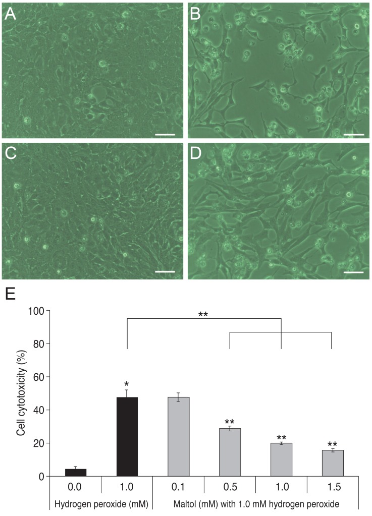
R28 cells were exposed to 0.0, 0.2, 0.4, 0.6, 0.8, 1.0, and 1.5 mM H2O2 for 24 hours and exposed to 1.0 mM H2O2 for 1, 3, 6, 16, 24 and 48 hours. Cytotoxicity was quantified by measurement of released LDH to culture media from injured cells. As shown in Fig. 1, H2O2 increased LDH release in a dose- and time-dependent manner. When R28 cells were exposed to 1.0 mM H2O2 for 24 hours, the cytotoxicity was 60.69 ± 5.71%. The 24 hours of exposure to 1.0 mM H2O2 was selected as the oxidative stress conditions for further experiments.
Effect of maltol against oxidative stress-induced cell damage on R28 cells
R28 cells were co-cultured with 1.0 mM H2O2 and different concentrations of maltol ranging from 0.1 to 1.5 mM for 24 hours. Co-culture with maltol attenuated H2O2-induced cytotoxicity in a dose-dependent manner (Fig. 2). When compared to 1.0 mM H2O2 treated cells, the protective effect of 0.1 mM maltol was not statistically different, but 0.5, 1.0 and 1.5 mM maltol significantly decreased LDH leakage from injured cells. LDH leakage markedly increased with the addition of 1.0 mM H2O2 (59.25 ± 2.81%) when compared to a control (10.48 ± 1.80%, p < 0.05). However, 1.0 mM maltol treatment significantly attenuated these cytotoxic effects (19.86 ± 1.11%). To investigate the underlying mechanism of the neuroprotective effects of maltol, the co-culture condition of 1.0 mM H2O2 and 1.0 mM maltol was selected for following experiments.
Maltol attenuates oxidative stress-induced apoptosis of R28 cells
In Fig. 3, the TUNEL assay for broken DNA from apoptotic cells revealed a FITC (green fluorescence)-positive signal. Propidium iodide counter-staining demonstrated living cells (red fluorescence). After co-culture with 1.0 mM maltol, R28 cells became more resistant to H2O2-induced oxidative stress injury. The ratio of TUNEL positive apoptotic cells to the total number of cells increased with the addition of 1.0 mM H2O2 (16.33 ± 2.31%) when compared to a control (1.72 ± 0.31 %, p < 0.05). However, the ratio decreased with co-culture of 1.0 mM maltol (2.64 ± 0.52 %). Maltol treatment markedly reduced the loss of R28 cells possibly through a decrease in apoptosis.
The neuroprotective mechanism of maltol in oxidatively-stressed R28 cells
To investigate the neuroprotective mechanism of maltol, the expression of nuclear factor-kappa B (NF-κB), extracellular signal-regulated kinase (ERK), c-Jun N-terminal kinase (JNK) and p38 were evaluated by Western immunoblot analysis. Fig. 4A shows the protein expression of phospho-NF-κB, phospho-JNK, phospho-ERK, phospho-p38, and β-actin after 48 hours with or without 1.0 mM maltol in R28 cells exposed to 1.0 mM H2O2. The expression of phospho-NF-κB, phospho-JNK, and phospho-ERK decreased with co-culture of 1.0 mM maltol, especially after 2 hours of H2O2 exposure. However, the expression of phospho-p38 showed no difference in the presence or absence of maltol. To quantify protein expression within 2 hours after exposure to 1.0 mM H2O2, the band intensities were measured with the ImageJ program (ImageJ Software; National Institute of Health, Bethesda, MD, USA). Fig. 4B shows that the expression levels of phospho-NF-κB, phospho-JNK, and phospho-ERK were 2.16-, 7.7-, and 4.3-times higher than those of a control, and these expression levels were significantly decreased with the addition of 1.0 mM maltol to 1.18-, 2.3-, and 1.8-times higher than those of the control, respectively. However, there was not significant change in phospho-p38 expression in oxidatively-stressed R28 cells at 1.7-times higher levels than control without maltol and 1.8-times with maltol.
Discussion
Maltol has been reported to have various biologic functions such as being a natural scavenger of reactive oxygen species [6], and as a mediator of enhanced mitochondrial function, reduced intracellular calcium, and reduced expression of NF-κB [7]. However, its effects and underlying mechanisms of action in neurosensory retinal cells have not been previously reported. In the present study, maltol not only increased cell viability but also attenuated DNA fragmentation. The results here show that maltol has neuroprotective effects against hypoxia-induced neurosensory retinal cell damage in R28 cells and its effects may act through the NFκB and mitogen-activated protein kinase (MAPK) signaling pathways.
NF-κB is an important transcription factor that regulates the stimulus-dependent induction of genes critical to the survival and death of neurons [11,12]. Various stimuli such as stress and cytokines can activate NF-κB, and then the activated form of phosphorylated NF-κB enters the nucleus and initiates the expression of genes to determine cell survival or death [13,14]. Wu et al. [15] reported that in light-induced photoreceptor degeneration, NF-κB was activated and exhibited neuroprotective and anti-apoptotic functions. However, Grilli et al. [16] reported that in glutamate-induced neurotoxicity, blockade of NF-κB activation was found to be protective. Therefore, NF-κB activation may induce both anti- and pro-apoptotic effects on different cells or under different pathological conditions. Previously, the activated NF-κB has been reported to participate in glutamate-induced neurotoxicity, N-methyl-D-aspartate (NMDA)-induced retinal neuronal cell death, retinal ischemia, and reperfusion injury [16,17,18,19]. In this study, phosphorylated NF-κB was increased in oxidatively-stressed R28 cells by H2O2, but was decreased when co-cultured with maltol. This finding is consistent with the results from previous studies. Yang et al. [7] reported that maltol could reduce NF-κB activated by H2O2 and prevent H2O2-induced apoptosis in human neuroblastoma cells (SH-SY5Y). This result shows that the neuroprotective effect of maltol is mediated by reduction of activated NF-κB.
The MAPK family consists of three main protein kinase families: the ERKs, JNKs, and p38 family of kinases. MAPK pathways have crucial roles in the regulation of cellular activities including proliferation, differentiation, survival, death, and cellular responses to external stresses. Activation of ERKs typically contributes to cell differentiation, proliferation, and survival, whereas JNK and p38 are, in contrast, activated by pro-inflammatory cytokines and environmental stress and have been suggested to promote apoptosis.
The present study showed that there were significant changes of phosphorylated ERK in oxidatively-stressed R28 cells with or without maltol treatment. Previous studies reported that ERK activation is a major factor related to cell survival [20,21,22]. However, in the present study, activated phosphorylated ERK decreased with maltol treatment in oxidatively-stressed R28 cells. The present findings are in apparent contrast to the above-mentioned studies. However, Luo et al. [23] demonstrated that inhibition of the ERK pathway promotes RGC survival and axon regeneration, probably via preventing excitotoxic amino acid release or blockade of nonapoptotic processes after optic nerve injury. Additionally, Cagnol et al. [24] reported that prolonged activation of the ERK pathway induces activation of pro-apoptotic caspase-8, and Stanciu et al. [25] reported that ERK activation is involved in glutamate and NMDA receptor-mediated neurotoxicity. The current study does not provide enough evidence to clarify the controversial role of the ERK pathway, because ERK activation could be interpreted as having both anti- and pro-apoptotic effects. Maltol may have a neuroprotective effect through down-regulation of phosphorylated ERK as a pro-apoptotic signal, or maltol may sufficiently reduce the oxidative stress on RGCs such that ERK cannot be activated as an anti-apoptotic signal. Although the role of phosphorylated ERK has not been clarified, the neuroprotective mechanism of maltol has been found to be associated with the ERK pathway in oxidatively-stressed R28 cells.
In this study, phosphorylated JNK was increased in oxidatively-stressed R28 cells and decreased with maltol addition. Previous studies demonstrated that oxidative stress-induced apoptosis was commonly mediated by the activation of JNK pathways [26]. The present study also showed that H2O2 induced activation of JNK and was suppressed by maltol addition. These results suggest that maltol may suppress the pro-apoptotic effect mediated by downregulation of phosphorylated JNK. On the other hand, there was no significant change in phospho-p38 in oxidatively-stressed R28 cells in the presence or absence of maltol. In the present study, phospho-p38 was found to increase less than phospho-ERK and JNK, at 1.7-, 7.7-, and 4.3-times higher than a control, respectively. Although p38 and JNK are often coactivated, specific activation of p38 and JNK has been observed, implying that there is specific activation of p38 or JNK. For example, Ogura and Kitamura [27] demonstrated that treatment of macrophages with menadione rapidly induced phosphorylation of ERK and p38 MAP kinase, but not JNK. The present study also showed that in oxidatively-stressed R28 cells, the p38 pathway was less influenced than other MAPK pathways.
R28 cells have both glial and neuronal characteristics, and have been widely used to elucidate the molecular mechanism of neuronal apoptosis in the retina [28]. However, the RGCs comprise only a very small fraction of retinal cells (one per ten thousand) and R28 cells do not behave like primary RGCs in every aspect. This is a potential limitation of the present study. However, isolating and culturing primary RGCs are technically difficult and widely used RGC-5 cells, once thought to be a rat RGC line [29], are now reported to express photoreceptor markers that are also expressed in the unrelated SV40-transformed mouse photoreceptor cell line 661W [30,31]. Use of this crosscontaminated and uncharacterized cell line could result in erroneous scientific conclusions. Therefore, the goal of this study was to use the R28 cell line to determine the neuroprotective effect of maltol with the expectation that the in vitro data set obtained from R28 cells can be verified in primary RGCs in a future study. Further, an in vitro cell line that is easy to handle and better represents RGC characteristics should be developed.
In this study, maltol was found to have a neuroprotective effect against H2O2-induced cytotoxicity in R28 cells. However, maltol has been reported to have weak mutagenic activity and toxic effects mediated through apoptosis [32]. However, at the recommended concentration, maltol does not have any toxic effect. The UN Joint FAO/WHO Export Committee on Food Additives concluded that up to 1 mg/kg per day was an acceptable level of consumption for human beings [2]. For dogs and rats, maltol showed no adverse toxic effect at doses up to and including 200 mg/kg/day for 2 years [33]. Also, Kim et al. [6] reported that maltol had a neuroprotective effect against oxidative stress in the brains of mice challenged with kainic acid at a dose of 100 mg/kg for 5 consecutive days. Previous in vitro studies showed that concentrations ranging from 50 to 1,000 µM did not significantly affect cell viability, but a concentration of 10 mM decreased cell viability to 75% [34]. Based on these findings, the present studies were conducted at concentrations of maltol ranging from 0.1 to 1.5 mM.
In conclusion, the present study demonstrates that maltol inhibits apoptosis of R28 cells induced by H2O2. The mechanisms underlying this protection are not fully understood but are associated with NF-κB and MAPK-related pathways. This study provides evidence that maltol could be an innovative neuroprotective therapeutic agent for the reduction of neurosensory retinal cell injury related to hypoxia.


 PDF Links
 PubReader
 Full text via DOI
 Full text via PMC
 Download Citation
 Print
Print


