

Dear Editor,
Mucopolysaccharidosis is an inheritable storage disease with deficiency of lysosomal enzymes that degrade glycosaminoglycans [1]. Mucopolysaccharidosis type II, also known as Hunter syndrome, is an X-linked inherited deficiency of enzyme iduronate 2-sulfatase, resulting in progressive accumulation of dermatan sulfate and heparin sulfate [1]. In many mucopolysaccharidoses, glycosaminoglycans accumulate in the retinal pigment epithelial (RPE) cells, resulting in pronounced lysosomal distension [2]. Previous studies have revealed outer nuclear layer thinning and the absence of outer segments, as well as reduced inner segments from the anterior midperipheral retina [2,3]. However, structural changes of the retina determined by high-resolution spectral domain optical coherence tomography (SD-OCT; Spectralis, Heidelberg Engineering, Heidelberg, Germany) in the early stage of Hunter syndrome with visual impairment have not been reported. With the introduction of SD-OCT, high-resolution images of the retina and optic disc could reveal occult and early structural changes related to disease progression [4]. Herein, we report a patient with Hunter syndrome whose visual acuity decrease was associated with early outer retinal layer changes found on SD-OCT.
A 5-year-old boy with Hunter syndrome was referred to our clinic for evaluation of ocular involvement. At initial presentation, his best corrected visual acuities were 20 / 25 in both eyes (OU). Cycloplegic refractive errors were +4.50 Dsph = -3.00 Dcyl ├Ś axis 180 in the right eye (OD) and +4.50 Dsph = -3.75 Dcyl ├Ś axis 180 in the left eye (OS). Slit lamp biomicroscopy showed clear corneas OU. Funduscopic examination was unremarkable.
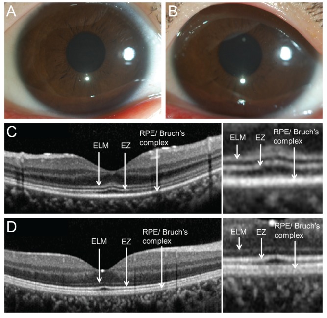
At the age of 9, he complained of visual acuity decrease OU. On ophthalmic examination, best corrected visual acuities were 20 / 30 OD and 20 / 50 OS. Cycloplegic refractive errors were +4.00 Dsph = -3.50 Dcyl ├Ś axis 180 OD and +4.00 Dsph = -4.25 Dcyl ├Ś axis 180 OS. Slit lamp biomicroscopy showed clear corneas OU (Fig. 1A and 1B). There was no relative afferent papillary defect. Optic disc margins showed mild blurring. However, lumbar spinal tapping revealed normal intracranial pressure. Visual field testing with Goldmann kinetic perimetry were normal OU. On funduscopic examination, the retina looked grossly intact, but standard electroretinography revealed a reduction of scotopic responses, consistent with rod cell degeneration. Reduced b-wave amplitudes in the maximal responses and reduced oscillatory potentials were also observed OU.
SD-OCT showed thickening of the external limiting membrane at the central macula, especially near the fovea (Fig. 1C and 1D). The ellipsoid line formerly known as the inner and outer segment photoreceptor junction appeared normal in thickness and contour, well distinguished from the RPE layer underneath. The peripapillary circular scan images showed a mild increase in bilateral peripapillary retinal nerve fiber layer thickness.
Our study demonstrates the structural changes of the retina determined by high-resolution SD-OCT in the early stage of Hunter syndrome. Previously, Yoon et al. [3] reported high-speed, ultrahigh-resolution OCT findings of the retina in Hunter syndrome. Stratus OCT revealed a loss of photoreceptors outside the fovea and cystoid spaces within the inner nuclear layer, ganglion cell layer, and outer nuclear layer [3]. Our patient only showed thickening of the external limiting membrane with no other abnormal findings in the RPE, suggesting that changes of the outer nuclear and photoreceptor layers precede RPE abnormalities. In contrast to our study, the previous case experienced more variable ocular symptoms typical of Hunter syndrome, such as exophthalmos, hypertelorism, and bilateral visual field loss with a ring scotoma pattern, which were not found in our patient.
The external limiting membrane, between photoreceptor inner segments and M├╝ller cell apical processes, is filled with the interphotoreceptor matrix, which contains a heterogeneous collection of glycoproteins, enzymes, and glycosaminoglycans [5]. Our SD-OCT findings correlate well with previous histopathological findings in the early stage of Hunter syndrome. The typical histopathological findings reported previously, such as a total loss of RPE, an overall loss of rods and cones, and reduced outer nuclear layer with atrophic bipolar cells, are usually shown as the disease progresses [2].
In conclusion, SD-OCT can be used as a diagnostic modality to monitor patients with Hunter syndrome and to detect subclinical progression of the disease in the early stages.
 PDF Links
 PubReader
 Full text via DOI
 Full text via PMC
 Download Citation
 Print
Print


