

The retinal pigment epithelium (RPE) plays an essential role in maintaining the viability and functionality of the neural retina and, among other functions, prevents the neurosensory retina from accumulating extracellular fluid in the subretinal space [1].
The human RPE cell line (ARPE-19) has structural and functional properties characteristic of RPE cells in vivo and therefore makes this cell line valuable for in vitro studies of retinal pigment epithelium physiology. These cells were first described as developing into highly differentiated, polarized monolayers and exhibiting features of a polarized and differentiated epithelium [2,3]. The measurement of transepithelial resistance (TER) in cultured ARPE cells has been used to evaluate the barrier function of RPE cells [4,5].
Adrenergic ╬▒ and ╬▓ receptors have been identified in cultured human RPE cells; they participate in membrane permeability and membrane potential in ARPE cells [6]. Also, it has been reported that ╬▒-2-receptors are expressed in RPE-choriocapillaries, though the exact function of this ╬▒ receptor has not yet been elucidated [7].
Brimonidine is a selective ╬▒-2-adrenergic agonist that has recently been approved for the long-term treatment of elevated intraocular pressure due to open angle glaucoma or ocular hypertension. In addition, it reaches significant concentrations with clinical dosing and exhibits a neuroprotective effect in animal models [8].
In the present study, we measured the TER in cultured ARPE cells in order to investigate the role of adrenergic ╬▒-2-agonists with respect to barrier function in the RPE.
Materials and Methods
Cell culture
The human RPE cell line, ARPE-19 cells were purchased from the American Type Culture Collection (Rockville, MD, USA). The cells were passaged and propagated using a slight modification of the technique described by Dunn et al [2]. In brief, the cells were cultured in medium (DMEM/F12; Gibco BRL, Grand Island, NY, USA) containing 10% fetal bovine serum (FBS; ThermoTrace, Melbourne, Australia) in a humidified incubator at 37Ōäā in 5% CO2. Media were changed twice a week.
The ARPE cells were seeded at a density of 1.66├Ś105 cells/cm2 in DMEM/F-12 culture medium and supplemented with 100 U/mL penicillin-streptomycin, 2 mM L-glutamine, and 1% FBS on a microporous filter (Transwell; Costar Corning, Corning, NY, USA) until the cells cultured to confluence.
For the hypoxic cell group, the ARPE-19 cells were transferred to a hypoxic chamber containing 5% CO2, 94% N2 and 1% O2 24 hours prior to TER measurement.
Measurement of transepithelial electrical resistance
The TER was measured using an epithelial volt-ohmmeter (EVOM; World Precision Instruments, Sarasota, FL, USA). The TER of the filter alone was measured as background and was subtracted from the TER obtained with the filters and ARPE-19 cells. Net TER measurements were calculated by subtracting the value of a blank, laminin-coated filter without cells from the experimental value. Final resistance-area products (╬®├Ścm2) were obtained by multiplication with the effective growth area.
The ARPE cells were cultured and the TER was measured every week for up to six weeks in order to determine the optimal incubation time for yielding the highest TER. The viability of the cells was assessed using trypan blue dye.
Brimonidine was purchased from Sigma Chemicals (St Louis, MO, USA) and was dissolved in dimethyl sulfoxide (DMSO) to a concentration of 50 mM and diluted to 0.01, 0.1, or 1 uM in DMEM/F12 medium containing 1% FBS just prior to use. The baseline TER was measured before administration of brimonidine. Immediately following this, 50 uL brimonidine solution was added to the apical chamber, and 50 uL culture media was taken out of the apical chamber simultaneously so that the concentration of brimonidine in the apical chamber reached 1, 10, or 100 nM (depending on the initial concentration) and so that the fluid volume of the apical chamber was not altered. To determine the optimal concentration of brimonidine, changes in TER were measured every 10 seconds for the first minute following exposure to each concentration of brimonidine and at 2, 3, and 5 minutes thereafter. Measurements were repeated at least three times for each well and for five wells of each concentration.
Changes in TER following administration of brimonidine to both the normoxic and hypoxic cell cultures were measured at every 10 seconds for the first minute following exposure and at 2, 3, 5, 10, 20, 30, and 120 minutes thereafter. Measurements were repeated at least three times for each well, and each experiment was repeated in 11 different wells.
Statistical analysis
The results were expressed as mean value┬▒SD. Comparison of the TERs between the normoxic and hypoxic group was performed using the Mann-Whitney U-test. Changes in the TER from the baseline value following treatment with brimonidine were analyzed using the Wilcoxon signed rank test. Statistical significance was accepted at the p<0.05 level. Statistical analyses were performed using SPSS ver. 17.0 (SPSS Inc., Chicago, IL, USA).
Results
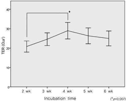
The monolayer of cells developed a resistance of approximately 20.9┬▒5.40 ╬®cm2(n=5) after two weeks of culture and reached a peak resistance of 29.1┬▒7.97 ╬®cm2 (n=5) after four weeks of culture. The TER had decreased to 25.1┬▒7.21 ╬®cm2 (n=5) after six weeks of culture (Fig. 1). The viability of the ARPE cells was greater than 97% at all measuring points.
When the ARPE monolayer was treated with various concentrations of brimonidine at four weeks of culture, the 10 nM concentration of brimonidine induced the greatest change in TER, although it was not statistically significant.
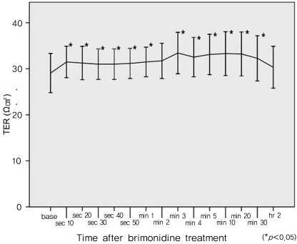
The normoxic ARPE monolayer was treated with a 10 nM concentration of brimonidine at four weeks of culture; this was believed to be the optimal concentration and culture period for TER measurement. Treatment with brimonidine resulted in an increase in TER (Fig. 2). Pretreatment values of TER (mean 29.1┬▒7.97 ╬®cm2) increased to 31.5┬▒6.0 ╬®cm2 1 minute after treatment (p=0.05, n=11, Wilcoxon signed rank test) and maintained this level 30 minutes (32.3┬▒9.16 ╬®cm2, p=0.038) after exposure. Thereafter, the TER decreased until it returned to its baseline level two hours following brimonidine treatment.
Discussion
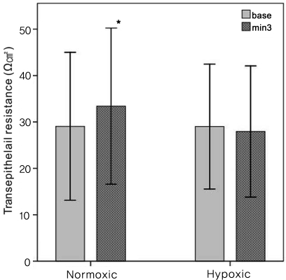
This study indicates that the TER of an ARPE-19 monolayer can be increased by treatment with a nanomolar amount of apical brimonidine. However, this effect was not reproduced in the ARPE monolayer treated under hypoxic conditions. These results suggest that ╬▒-2 receptors play a role in maintaining barrier function in the RPE.
The RPE passively and actively transports fluid in the subretinal to choroidal direction; this RPE fluid "pump" function is thought to play a major role in reabsorption of subretinal fluid and in promoting and maintaining retinal attachment [9]. Therefore, the RPE membrane is a potential target for small therapeutic molecules that bind membrane receptors and activate fluid absorption from the subretinal space.
Several studies have reported that ╬▒-2-adrenergic receptors are present in the human ciliary body, retinal pigmented epithelium-choriocapillaris, iris, and neurosensory retina [10,11]. Quinn and associates reported that adrenergic agonists, including isoproterenol and epinephrine, activate ion transport at the apical membrane of the human fetal RPE [6]. We hypothesize that the presence of ╬▒-2-receptors in the RPE could change the barrier function of the RPE membrane.
Brimonidine, an ╬▒-2-adrenergic receptor agonist, has been employed in the clinical setting of glaucoma because of its therapeutic potential for reducing intraocular pressure (IOP) by aqueous suppression and enhanced uveoscleral outflow. In addition to these IOP-lowering effects, several recent investigations in animal models suggest that brimonidine may exert a neuroprotective effect on retinal ganglion cells by preventing cell death secondary to ischemic injury [8,12]. A recent clinical pilot study suggests a trend toward slower progression of retinal dystrophy when treated with topical brimonidine [13]. Furthermore, topically applied brimonidine can reach the back of the eye at nanomolar concentrations sufficient to activate ╬▒-2 adrenergic receptors. This yields obvious advantages for the treatment of various retinal diseases in clinical settings [14].
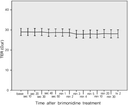
Following exposure to brimonidine, there was no increase in the TER of the ARPE membrane culture subjected to hypoxic conditions. This result suggests that hypoxia can induce dysfunction of active ion transport at the apical membrane of the ARPE.
Some studies have reported that DMSO at concentrations of 1% or 10% can induce TER changes in various cell membranes [15,16]. In contrast, Cui et al. [5] showed that 0.05% DMSO did not alter TER in cultured ARPE. In this study, the final concentration of DMSO was 1/250 of that used in the former study, and its effect on TER was therefore insignificant
One limitation of this study is that we could not elucidate the mechanisms of TER change in the ARPE membrane. An analysis of ion transfer and permeability testing could have possibly supported the TER changes observed in this study.
Although the exact mechanism remains unclear, brimonidine enhanced the barrier function in ARPE-19 cells in the present study. The specific effects of brimonidine on the action of ion pumps require further investigation.
 PDF Links
 PubReader
 Full text via DOI
 Full text via PMC
 Download Citation
 Print
Print


